Organisation
Work through one section at a time. Higher-only material is labelled explicitly; optional challenges can also use Foundation content.
 GCSE BIOLOGY · AQA TRILOGYTutor dashboardSaves on this device
Your answers save automatically as you work.
Saved in this browser on this device.
Discover how tissues, organs and transport systems work together in humans and plants, and how evidence helps us understand health and disease.
Supported GCSE version · Clear examples, worked calculations and optional challenges.
Work through one section at a time. Higher-only material is labelled explicitly; optional challenges can also use Foundation content.
Cells with similar structures working together on a shared function.
Different tissues working together, such as the stomach.
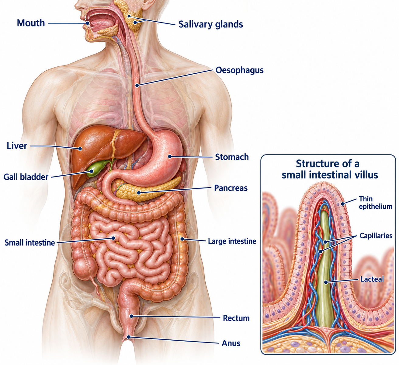
Organs cooperating in a major function, such as digestion.
A protein catalyst that speeds up a biological reaction without being used up.
The molecule an enzyme acts on.
The shaped region of an enzyme where its substrate binds.
An enzyme whose shape has changed so its active site no longer works properly.
Breaking large fat globules into small droplets; this increases surface area.
Movement of digested soluble molecules through the intestine wall into the body.
The liquid part of blood that carries cells and dissolved substances.
An artery supplying the heart muscle itself.
Something associated with a greater chance of developing a disease.
An association between variables; it does not alone establish cause.
Describes a cancerous tumour able to invade and spread.
Loss of water vapour from a plant, mainly through leaf stomata.
Transport of dissolved sugars through phloem.
A pore in the leaf surface, controlled by guard cells; plural: stomata.
Plant growth tissue containing cells that divide and differentiate.
The space inside a tube such as a blood vessel.
A test identifying whether a substance is present rather than measuring an exact amount.
A cell is the smallest living unit. A tissue contains cells with similar structures working together. An organ contains different tissues working together. An organ system contains organs that carry out a major body function.
For example: a muscle cell → muscle tissue → stomach → digestive system → human organism. The stomach also contains glandular tissue that makes digestive substances and epithelial tissue that covers surfaces. One organ needs several tissues because it has several jobs.
Large food molecules cannot pass easily through the gut wall. Digestion breaks them into small, soluble molecules. Absorption is their movement through the small-intestine wall into the body. These are different processes.
Absorbed sugars, amino acids and lipid products provide raw materials for growth and repair. Cells use them to build new carbohydrates, proteins and lipids. Glucose also releases energy through respiration. These chemical reactions form part of metabolism.
An enzyme is a protein that acts as a biological catalyst. It speeds up a reaction without being used up. A substrate is the molecule an enzyme acts on. The substrate binds at a specially shaped region called the active site.
In the simplified lock-and-key model, only a substrate with a matching shape fits. An enzyme therefore catalyses a specific reaction. Products leave the active site, allowing the enzyme to be used again.
In cooler conditions, particles move more slowly and collide less often, so the reaction is slower. Warming towards the optimum temperature increases successful collisions. Above the optimum, bonds maintaining the enzyme's shape are disrupted. Its active site changes shape, so the substrate no longer fits well. This is denaturation.
Each enzyme also has an optimum pH. Moving far from it can change the active-site shape. A stomach protease works in acidic conditions; many small-intestine enzymes work best in alkaline conditions.
The substrate fits the active site and is converted into products. The enzyme is unchanged and can work again. High temperatures change the active site shape so the substrate no longer fits.
Model limitations: The protein and molecules are schematic, not accurate molecular structures. The 55 °C denaturation threshold is chosen for this model; real enzymes vary and denaturation also depends on exposure time. This shows a breakdown reaction, not every enzyme reaction.
| Enzyme | Made in | Reaction |
|---|---|---|
| Amylase, a carbohydrase | Salivary glands, pancreas, small intestine | Starch → smaller sugars |
| Proteases | Stomach, pancreas, small intestine | Proteins → amino acids |
| Lipases | Pancreas, small intestine | Lipids → fatty acids + glycerol |
Carbohydrases as a group break carbohydrates down into simple sugars. At this level, use the word equations above; digestive chemical symbol equations are not required.
Bile is not an enzyme. It is made by the liver, stored in the gall bladder and released into the small intestine. It is alkaline, so it neutralises acid arriving from the stomach. It also emulsifies fat: large fat globules become many small droplets. This provides more surface area for lipase to act on. Emulsification does not itself digest fat molecules.
These are qualitative tests: they identify whether a substance is present. Prepare food extracts by crushing solid food with distilled water and filtering if needed. Use separate clean samples for each test. Wear eye protection, avoid skin contact with reagents and never taste food in a laboratory.
| Substance | Method | Positive result |
|---|---|---|
| Reducing sugars | Add Benedict's solution, then heat in a hot water bath. | Blue changes through green/yellow/orange towards a brick-red precipitate, depending on concentration. |
| Starch | Add iodine solution. | Orange-brown becomes blue-black. |
| Protein | Add Biuret reagent. | Blue becomes lilac/purple. |
| Lipids | Add Sudan III solution and mix gently, following the supplied method. | A distinct stained red oil layer forms. |
An alternative lipid test is the ethanol emulsion test: shake the sample with ethanol, then add the extract to water. A cloudy white emulsion indicates lipid. Ethanol is flammable: use no naked flame. Keep hot water baths away from ethanol.
Use a known positive sample to check the reagent works and water as a negative control. Compare samples using the same volumes and conditions. A negative result means the substance was not detected; very small amounts may be below the test's detection limit.
Question: how does pH affect the time amylase takes to digest starch? Use solutions prepared at different known pH values.
The independent variable is pH; the measured dependent variable is time until starch is no longer detected. The water bath controls temperature. Use fresh pipettes or wash them to prevent contamination. Take care with hot water and iodine.
For equal amounts of starch, an estimated rate is 1 ÷ time. If time is 60 s, rate = 1/60 = 0.0167 s⁻¹. A shorter time means a faster reaction.
Plot pH on the horizontal axis and rate on the vertical axis. Do not assume every enzyme has an optimum of pH 7: use the evidence supplied.
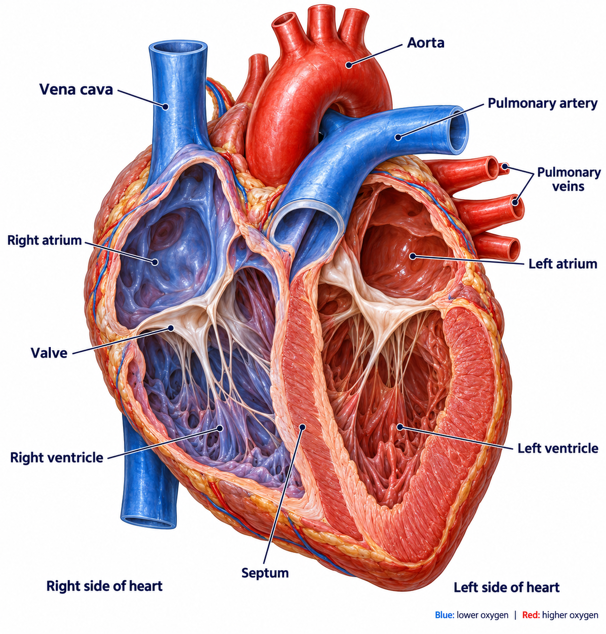
The heart is a muscular pump. In a double circulatory system, blood passes through the heart twice during one complete journey around the lungs and body.
Body → vena cava → right atrium → right ventricle → pulmonary artery → lungs → pulmonary vein → left atrium → left ventricle → aorta → body.
The right ventricle pumps blood to the nearby lungs. The left ventricle has a thicker muscular wall because it must produce higher pressure to supply the whole body. Valves prevent backflow. The septum separates the left and right sides so oxygenated and deoxygenated blood do not mix.
The coronary arteries supply the heart muscle itself with oxygen and glucose for respiration. Blood inside the chambers does not replace this supply.
A group of cells in the right atrium acts as the heart's natural pacemaker. Electrical signals coordinate the heartbeat. An artificial pacemaker supplies electrical impulses to help control an abnormal heart rhythm.
For flow calculations, use volume per unit time. If a heart pumps 70 cm³ per beat at 72 beats per minute, blood flow = 70 × 72 = 5,040 cm³/min, or 5.04 dm³/min. Remember: 1,000 cm³ = 1 dm³.
At rest: 70 beats/min × 70 cm³/beat = 4,900 cm³/min. During exercise: 120 × 100 = 12,000 cm³/min. Flow increases by 7,100 cm³/min. Increased oxygen and glucose delivery supports more respiration in working muscles.
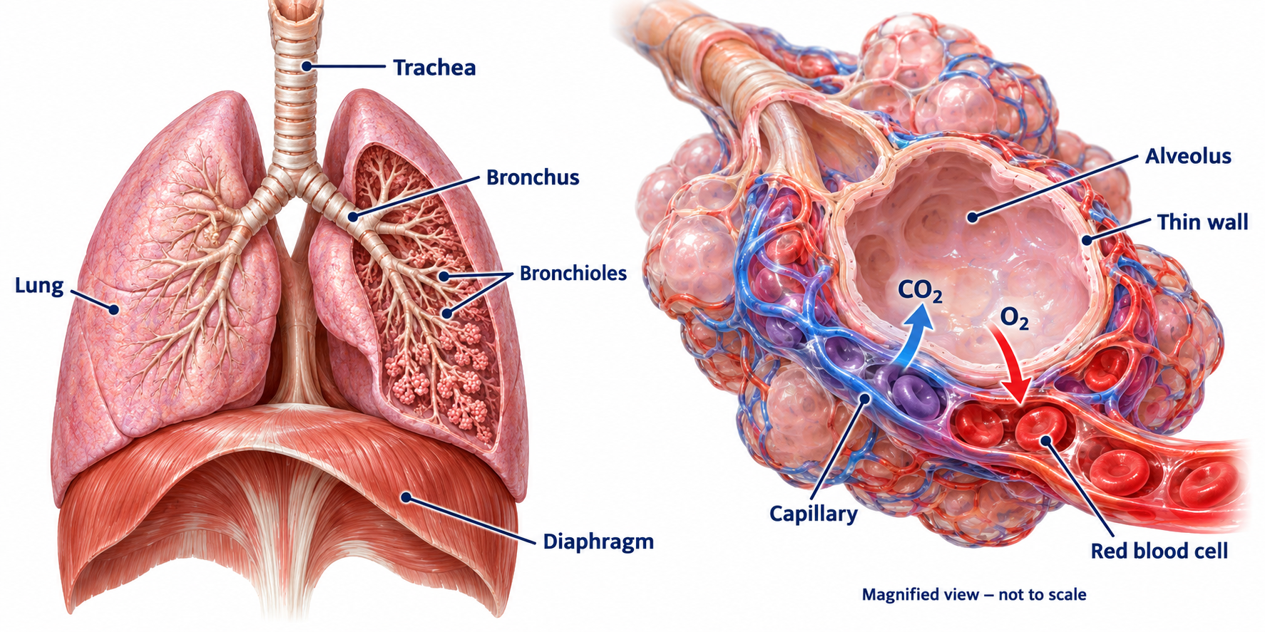
Air travels down the trachea, into the two bronchi and through smaller branches to the alveoli, the tiny air sacs. A capillary network surrounds each alveolus.
Oxygen moves by diffusion from alveolar air into blood. Carbon dioxide diffuses in the opposite direction. The many alveoli provide a large surface area. Alveolar and capillary walls are thin, giving a short diffusion distance. Breathing refreshes alveolar air and blood flow carries oxygen away, maintaining steep concentration gradients.
Gas exchange and respiration are different. Gas exchange moves gases between air and blood. Respiration is a set of chemical reactions in cells that release energy.
| Vessel | Structure | How it helps |
|---|---|---|
| Artery | Thick muscular, elastic wall; relatively narrow lumen | Withstands high pressure. Elastic recoil helps maintain blood flow between heartbeats. |
| Vein | Thinner wall, large lumen and valves | Allows low-pressure blood to return to the heart; valves prevent backflow. |
| Capillary | Wall only one cell thick; very narrow lumen; extensive networks | Short diffusion distance and large total area for exchange with tissues. |
A lumen is the space inside a vessel. Arteries do not all carry oxygenated blood. Their name describes their direction relative to the heart.
Blood contains a liquid called plasma with cells and cell fragments suspended in it.
In an image, look for numerous disc-shaped red cells, larger nucleated white cells and very small platelet fragments. Blood products can be life-saving, but compatibility and screening matter because immune reactions and infection transmission are possible risks.
In coronary heart disease, fatty deposits narrow coronary arteries. Less blood reaches heart muscle, so it receives less oxygen. Aerobic respiration falls; severe blockage can damage or kill heart muscle.
| Treatment | Possible benefit | Limitation or risk |
|---|---|---|
| Stent | A mesh tube holds a narrowed artery open and improves flow. | Insertion is a medical procedure; complications include clotting. It does not remove all underlying risk factors. |
| Statin | Lowers blood cholesterol and slows the build-up of fatty deposits. | Usually requires continued use; side effects can occur. It does not instantly reopen a blocked artery. |
| Replacement valve | Restores effective one-way flow when a valve leaks or does not open fully. | Surgery has risks. Mechanical valves can last longer but often require anti-clotting medication; biological valves can wear out. |
| Heart or heart-and-lung transplant | Replaces severely failing organs. | Donors are limited; surgery, rejection and immunosuppressant treatment have risks. |
| Artificial heart | Can support circulation while awaiting transplant or during recovery. | Mechanical complications, infection and clotting are possible. |
Choose a treatment by weighing the problem, likely benefit and risks for the patient. A pacemaker corrects rhythm; a stent opens an artery. They solve different problems.
Health includes physical and mental well-being. A person can have no diagnosed infection but still have poor health because of stress, diet or difficult living circumstances.
Communicable diseases can spread between organisms, usually through pathogens. Non-communicable diseases do not spread by infection, although infection can contribute to some of them.
Illness can affect family life, employment and independence. Communities and nations bear costs through healthcare, caring responsibilities and lost productivity. For example, a person may lose income or independence, relatives may provide unpaid care, local services may face greater demand, and national health systems may pay for long-term treatment. Comparing local, national and global patterns also requires considering differences in age, access to healthcare and data collection. These impacts are not just financial.
A risk factor is associated with a greater chance of disease. It does not guarantee that someone will develop it. Multiple factors can interact, including inherited factors, age, environment and lifestyle.
A diet high in saturated fat can raise blood cholesterol and contribute to fatty artery deposits. Regular physical activity helps maintain cardiovascular fitness and a healthy body mass. If energy intake regularly exceeds energy use, body fat can increase. Smoking damages blood vessels as well as lungs. These mechanisms help explain some associations; they do not make disease inevitable.
A correlation means two variables are associated. It does not by itself establish a cause. An observed link may involve a third variable, or confounding factor. Researchers look for repeatable patterns and plausible mechanisms, and compare groups while considering other differences.
Use large, representative samples. A survey of one sports club may not represent a whole town. Compare rates per equal population sizes, not just case totals.
Town A has 120 cases among 20,000 people: 120 ÷ 20,000 × 1,000 = 6 cases per 1,000. Town B has 80 cases among 10,000: 8 per 1,000. B has fewer cases in total but a higher rate.
Use a bar chart for distinct categories, such as smoker/non-smoker. Use a histogram for continuous data grouped into intervals, such as age. Use a scatter graph to investigate an association between two numerical variables. Give both axes labels and units where appropriate.
In a scatter graph, points rising from left to right suggest positive correlation. Falling points suggest negative correlation. A scattered pattern may show no clear correlation. Describe the pattern before explaining it.
Frequency tables organise counts. Include every group, label intervals without overlap and check that frequencies add to the sample size. When drawing histograms here, use equal-width intervals so frequencies can be compared directly.
Cancer develops when changes in cells lead to uncontrolled growth and division. A mass of abnormal cells is a tumour.
A benign tumour remains in one area and does not invade other tissues. It may still cause harm by pressing on structures. A malignant tumour is cancerous: cells invade neighbouring tissue and can travel in the blood to establish secondary tumours elsewhere.
Risk factors include inherited genetic variants and lifestyle or environmental exposure to carcinogens. A risk factor increases probability; it is not proof of why one individual developed cancer.
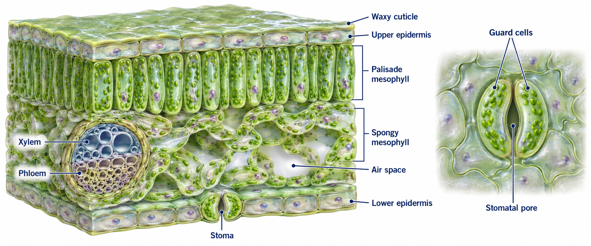
A leaf contains several tissues that work together to carry out photosynthesis and exchange gases.
Meristems at root and shoot tips contain cells that divide and can differentiate into new plant tissues. Meristems enable growth; they are not a mature leaf layer.
Roots, stems and leaves work as an organ system. Root hair cells have a long extension giving a large surface area. Water enters by osmosis; mineral ions can enter by active transport, using energy from respiration when ions move against a concentration gradient.
Xylem vessels are continuous hollow tubes formed from dead cells. Thickened lignified walls strengthen the tubes and help keep them open. They carry water and dissolved mineral ions upwards from roots to stems and leaves.
Phloem contains elongated living cells joined end to end. Pores in the end walls let cell sap pass between cells. Movement of dissolved sugars through phloem is translocation. Sugars move from sources, such as photosynthesising leaves, to places where they are used or stored. Movement can be upwards or downwards in different tubes.
Transpiration is the loss of water vapour from the aerial parts of a plant, mainly leaves. Water evaporates from moist mesophyll cell surfaces into leaf air spaces. Water vapour then diffuses out through open stomata. This loss helps draw a continuous stream of water up xylem.
| Change | Usual effect and explanation |
|---|---|
| Higher temperature | Faster evaporation and diffusion, so water loss usually rises. |
| Higher humidity | Smaller water-vapour concentration difference between leaf and air, so loss falls. |
| More air movement | Removes humid air near the leaf, maintaining a steeper gradient, so loss rises. |
| Greater light intensity | Stomata usually open for carbon-dioxide uptake in photosynthesis, increasing water loss. |
These are usual trends when other factors are controlled. Severe water shortage can cause stomata to close, reducing water loss but also limiting carbon-dioxide entry.
A potometer estimates water uptake by following an air bubble along a narrow capillary tube connected to a leafy shoot. It estimates transpiration because most absorbed water replaces evaporated water, but some is used in growth and photosynthesis.
Distance rate = distance ÷ time. To find volume uptake, use volume = capillary cross-sectional area × distance. For a circular tube, area = πr². If r = 0.5 mm and distance = 40 mm, volume = π × 0.5² × 40 = 31.4 mm³. In 5 min, rate = 6.28 mm³/min.
For stomata, examine epidermal impressions with a microscope. Count stomata in several randomly selected fields of known area. Calculate stomatal density = number ÷ area, then a mean. Sample both surfaces and several leaves rather than choosing the most crowded field.
For a continuous variable such as light intensity, plot it on the horizontal axis and water-uptake rate on the vertical axis. Select a scale that uses the graph area effectively and include units.
Start with the 53 main questions. Higher-only questions are labelled. You can complete this topic over several sessions; your answers save as you go. Use a hint if you get stuck, then compare your work with an example answer. Your tutor can mark written answers.
A student mixes starch and amylase at a constant temperature. Every 30 seconds, a drop of the mixture is added to fresh iodine on a spotting tile.
At 30 and 60 seconds, the iodine turns blue-black. At 90 seconds, it stays orange-brown.
(a) Starch is present. (b) 90 seconds. (c) Digestion finished between the samples at 60 and 90 seconds; testing more frequently would give a more precise time.
A sample has these ages in years: 12, 14, 17, 21, 23, 28, 29, 31, 35, 38, 41, 43. On paper, group them into 10–19, 20–29, 30–39 and 40–49. Draw a histogram using equal-width intervals and touching bars.
10–19: 3; 20–29: 4; 30–39: 3; 40–49: 2. Total = 12. Horizontal axis: age / years; vertical axis: frequency. These data are invented for practice.
| Relative humidity / % | Mean uptake / mm³ min⁻¹ |
|---|---|
| 30 | 18 |
| 50 | 13 |
| 70 | 8 |
| 90 | 3 |
On paper, plot humidity horizontally and uptake vertically. Describe the pattern, then explain it using a concentration gradient.
Uptake decreases as humidity increases. More water vapour outside the leaf makes the difference between leaf air spaces and surrounding air smaller, reducing net diffusion and water loss. Water uptake is an estimate of transpiration, not a direct measurement of evaporated water.
When connected with your pupil code, your answers save online for Kevin to see. Click Submit to Kevin when you have finished. Downloads are optional.
Connect with your pupil code above to submit.
Use a scientific idea and a specific example in your reflection.